
GE LOGIQ xe Color Doppler Ultrasound Used in Good Working for Hospital Medical
GE LOGIQ xe Color Doppler Ultrasound in Good Working Device
► Brief Description
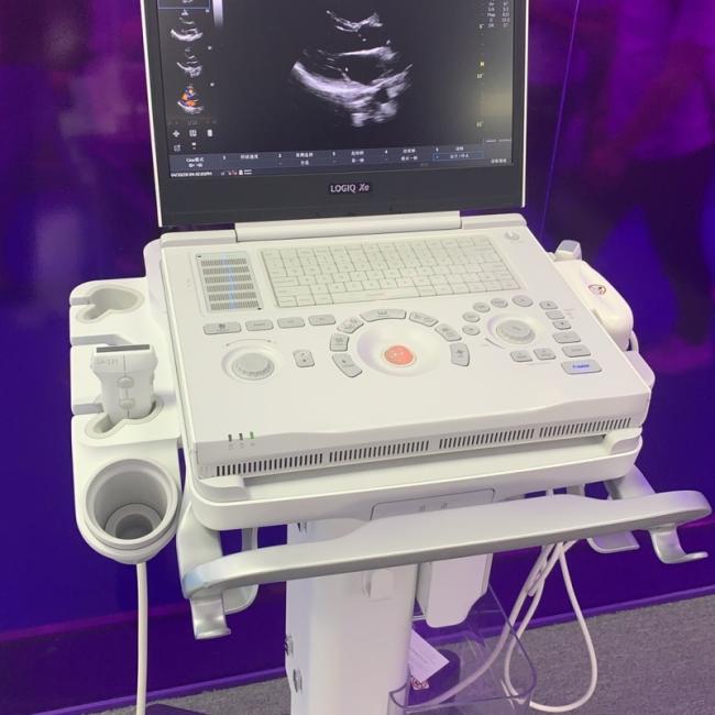
The GE LOGIQ xe is a portable color Doppler ultrasound diagnostic system. Weighing approximately 4.6 kg and measuring roughly 34 cm x 28.7 cm x 6.1 cm, its compact and lightweight design allows for easy mobility between departments. It features a 5-inch high-resolution color LCD display and three active probe ports that support seamless switching, accommodating various electronic array probes such as convex, linear, and phased array types.
In summary, this system is a battery-powered, portable ultrasound unit suitable for diverse clinical settings, including hospitals and medical clinics. It supports a wide range of imaging modes, including B-mode, M-mode, PW, CW, Color Doppler, Color M-mode, and Power Doppler. Beyond standard color Doppler capabilities, the system incorporates B-Flow™ technology, which provides real-time, high-resolution imaging of blood flow morphology, overcoming the limitations of conventional Doppler imaging.
► Specifications
| Product Name | Ultrasound |
| Model | LOGIQ xe |
| MOQ | 1 pc |
| Brand | GE |
| Payment Method | Paypal,T/T,Alipay |
| Warranty | 90 Days |
| Color | White |
| Delivery Method | DHL ,Fedex ,UPS |
| Stock Situation | Stock available |
| Delivery | 3-5 Working Days |
| Inventory | In stock |
| Use for | Hospital |
| Material | Plastic,Metal |
| Weight | 4.6KG |
►Detailed Photos
►Clinical Applications
1.Abdomen: Examination of organs such as the liver, gallbladder, pancreas, and kidneys.
2.Obstetrics and Gynecology: Ultrasound diagnosis of fetal conditions and gynecological disorders.
3.Cardiology: Assessment of cardiac structure and function in adults and children.
4.Vascular: Hemodynamic assessment of peripheral vessels, including those in the neck and limbs.
5.Superficial Organs: Examination of small organs such as the breast, thyroid, and testes.
6.Musculoskeletal (MSK): Diagnosis of disorders affecting joints, tendons, and muscles.
►GE LOGIQ xe Ultrasound Operating Procedures
1.Press the power button on the main unit to turn it on, and inspect the device and probe for any external damage or irregularities.
Insert the probe connector horizontally into the corresponding slot, then rotate the locking mechanism to the vertical position to secure it.
2.After entering patient information, the system automatically enters B-mode; the overall image brightness can be adjusted using the "B-Mode" knob on the control panel.
3.Press the "CF" (Color Flow) button on the control panel to activate Color Doppler mode. Rotate the "CF" knob to adjust the overall gain of the color signal for optimal sensitivity.
4.Optimize the image and measurements based on the target blood flow velocity: use high-pass filtering for high-velocity flow and low-pass filtering for low-velocity flow. If necessary, locate and activate the "Invert" option in the on-screen menu to reverse the color map.
When using a linear array probe, the "Steer" function allows you to angle the ultrasound beam to optimize the intersection angle between the blood flow and the beam.
5.Press the "Freeze" key to freeze the current image for detailed analysis.
Press the "Measure" key, use the trackball to position the measurement cursors, and confirm to display the measurement results on the screen.
Press the "P1" key to save the current image to the hard drive. To save a dynamic image sequence (Cine clip), press the "Mark Cine" key followed by the "P1" key.
If you want to explore more details for GE LOGIQ xe Color Doppler Ultrasound , please send the inquiry to me, Looking forward to hearing from you.
Get in Touch
Have questions about our products or want to discuss a custom order? Our team is ready to help you.