
Mindray Resona A20S Full body Application Color Doppler Ultrasound
Mindray Resona A20S Full body Application Color Doppler Ultrasound
Brief introduction
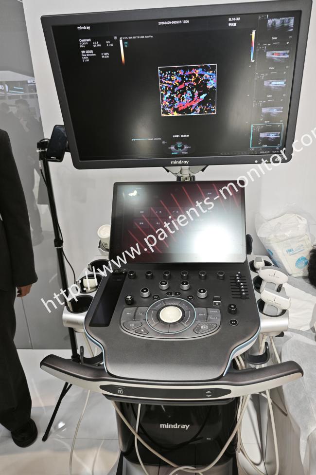
The Mindray Resona A20S is a premium, full‑body application color Doppler ultrasound system a super premium diagnostic platform that sits at the top of Mindray’s Resona A series. It is a general purpose diagnostic ultrasound system intended for use by qualified healthcare professionals for imaging across a wide range of clinical applications.
The system is built around Mindray’s Acoustic Wisdom platform, which integrates advanced sound‑electric data processing, imaging algorithms, and system computing power.
Functions
1. The 15.6-inch touchscreen supports gesture control and remembers recently used probe and mode combinations, allowing users to switch configurations with a single touch. The system supports wireless data transmission for convenient access to examination reports. Intelligent tracking and image optimization functions streamline the scanning process.
2. Utilizing advanced beamforming technology, the system delivers exceptional image quality. Its ultra-parallel computing architecture significantly enhances data processing capabilities, ensuring smooth real-time imaging.
3. AFM (Advanced Focusing Matrix) transducer technology significantly improves the sensitivity and bandwidth of acoustic and electrical signals, laying the foundation for image clarity. FreeBeam adaptive beamforming technology dynamically optimizes the main lobe and suppresses side lobes, providing higher resolution and contrast.
4. The system supports advanced interventional imaging and fusion imaging. It supports fusion imaging, synchronizing real-time ultrasound images with CT/MRI/ultrasound volumetric images. For liver interventional procedures, it provides preoperative 3D simulation and planning, as well as intraoperative 3D ablation guidance.
Routine operation
- Select the appropriate clinical application preset (abdominal, cardiac, vascular, obstetric, small parts, etc.).
- Select the appropriate transducer for the examination.
- Apply ultrasound gel to the transducer and begin scanning.
- Use the touchscreen or control panel to adjust imaging parameters as needed.
- For advanced functions – elastography, fusion imaging, 3D/4D – select the appropriate mode from the touchscreen.
- After the examination, save the images and measurements, generate the report, and export data as required.
Notice
Keep the system clean and follow infection control protocols. After each examination, clean the device surface and probes promptly to prevent cross‑infection. Use only approved cleaning agents that are compatible with the system materials.
Avoid strong electromagnetic fields. The system should be kept away from strong electromagnetic environments that could interfere with signals and cause measurement errors.
Ensure proper probe handling during examinations. Keep the probe perpendicular to the examination area and avoid excessive pressure that could distort the image. Select the appropriate probe type and frequency setting for each examination to obtain optimal image quality
Get in Touch
Have questions about our products or want to discuss a custom order? Our team is ready to help you.