
PHiliph Vivid E9 Color Doppler Ultrasound Diagnostic System
PHiliph Vivid E9 Color Doppler Ultrasound Diagnostic System
Introduction
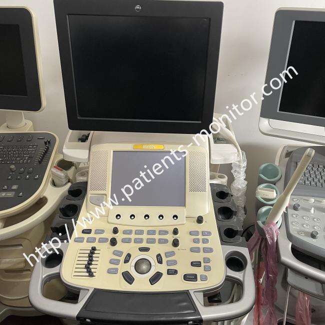
The Vivid E9 is a high‑performance cardiovascular ultrasound system a premium tier diagnostic platform built around an Accelerated Volume Architecture (AVA) with approximately eight times the processing power of conventional ultrasound systems.
The system is designed primarily for advanced cardiac imaging, with a particular emphasis on 4D (real time 3D) echocardiography. It supports single beat volume acquisition, which means you can capture a full volume dataset of the heart in a single cardiac cycle a practical advantage for patients with arrhythmias or irregular heart rhythms. The platform also includes advanced quantification tools such as 4D Stress, 4D Auto LV Quantification, and strain analysis.
Functions
1. Delivers true 4D cardiac imaging. The system's AVA architecture enables real‑time 4D volume imaging, allowing clinicians to visualise the heart in three dimensions over time. This is particularly useful for assessing valve morphology, ventricular function, and congenital heart defects.
2. Excels at myocardial quantification. The platform includes comprehensive tissue Doppler analysis : TVI, TTI, strain/strain rate, and TSI which are valuable for assessing regional wall motion and detecting subtle myocardial dysfunction.
3. Covers a broad range of applications. While the Vivid E9 is optimised for cardiology, it is a shared‑service system capable of abdominal, obstetric, gynaecological, vascular, and small parts imaging.
4. Supports advanced quantification tools. The system includes Auto IMT (intima media thickness measurement), AFI (automated function imaging), Scan Assist, and Flexi Volume tools that streamline workflow and reduce operator dependence
Operation:
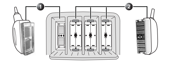
Probes can be connected at any time, whether the unit is on or off. The system has three types of probe ports; one PD probe port, three PDT probe ports and a Doppler probe port.
-
Select the appropriate transducer for the examination.
-
Apply ultrasound gel to the transducer and begin scanning.
-
Use the touchscreen or control panel to adjust imaging parameters as needed.
-
For 4D imaging, select the appropriate volume acquisition mode – the system can acquire a full volume in one, two, or multiple cardiac cycles.
-
After the examination, save the images and measurements, generate the report, and export data as required.
Please note
1. Keep the system clean and follow infection control protocols. After each examination, clean the device surface and probes promptly to prevent cross‑infection. Use only approved cleaning agents that are compatible with the system materials.
2. Avoid strong electromagnetic fields. The system should be kept away from strong electromagnetic environments that could interfere with signals and cause measurement errors.
3. Establish a reasonable service/maintenance plan. Regular maintenance is essential to prevent machine breakdown or personal injury. Service and repairs should only be performed by authorised service personnel.
Get in Touch
Have questions about our products or want to discuss a custom order? Our team is ready to help you.