
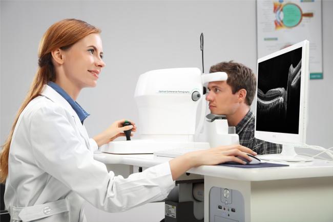
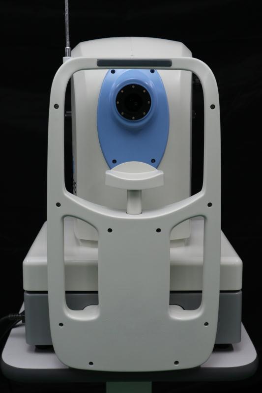
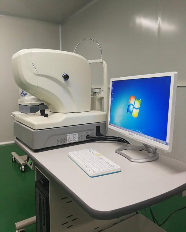
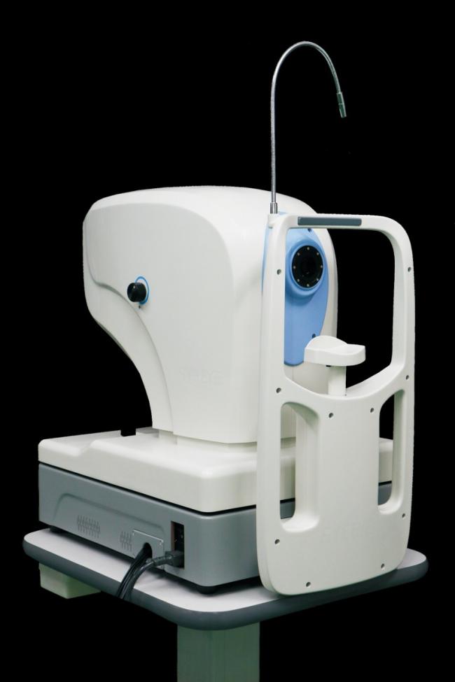
Professional Optical machine/ OCT with fundus camera function Optical Coherence Tomography
The OCT with fundus color photography with variable retina scanning modes and analysis procedures, which provides direct and reliable analysis reports for auxiliary examinations of retina diseases and glaucoma.
High- definition and comprehensive
High-definition OCT images with clear color fundus photography present a more accurate details in scanning area.
OCT scanning for macula
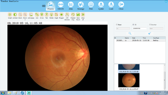
Fundus color photography
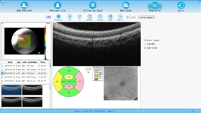
Analysis for macula thickness
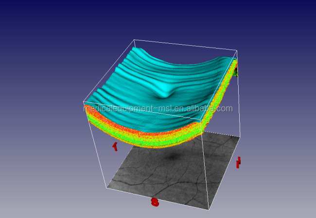
3D scanning in macula area of 6mm obtains more than 128 OCT images and mass data. And through professional calculated software, topographic map of retina thickness is got to analyze quality and quantity directly and precisely for diagnosis of retina diseases.
Analysis for macula lesion followup
Macula lesion follow-up visit analysis software helps doctors to judge progress of disease and effect of therapy, such as followup of age-related macula lesion, cystoid macular edema therapy comparison before and after, etc..
Get in Touch
Have questions about our products or want to discuss a custom order? Our team is ready to help you.